![]() The first digital mammography units arrived at the BCHS in 2012. Both units have now been classified by the vendor as end of product life in June 2021. This means that production of our current units has ceased.
The first digital mammography units arrived at the BCHS in 2012. Both units have now been classified by the vendor as end of product life in June 2021. This means that production of our current units has ceased.
Mammography is the most valuable tool radiologists rely on to diagnose breast cancer and to follow patients diagnosed with cancer post treatment. This specialized x-ray is used to produce detailed images of the breast. Breast tomosynthesis is an advanced form of mammographic imaging that allows for creation of 3 dimensional images of the breasts, proven to improve the ability of mammography to diagnose cancer in certain types of breast tissue.
Contrast enhanced mammography is an additional tool to aid in diagnosis of breast cancer. The new equipment at the BCHS will have both breast tomosynthesis and contrast enhanced mammographic capabilities.
Ultrasound imaging is used for a wide range of indications and symptoms and is also a complementary modality to mammography. It can help to diagnose breast lumps or other abnormalities found during an exam or on a mammogram.
Currently at BCHS, patients often have to travel back and forth between the mammographic suite and the ultrasound area when we are dealing with breast issues. The ideal future state is to create one area for breast imaging at the BCHS. Purchase of an additional ultrasound unit specifically for breast imaging will allow patients to remain in one location for all required breast imaging needs, including mammography, ultrasound, and biopsy, instead of being cared for throughout various areas of the department. This will also free up time in the general ultrasound area to take care of more inpatients and patients from the emergency department.

![]() The gamma camera is a machine that can detect and create images from very small amounts of ionizing radiation emitted from patients having a nuclear medicine exam.
The gamma camera is a machine that can detect and create images from very small amounts of ionizing radiation emitted from patients having a nuclear medicine exam.
With the existing gamma camera at end of life, BCHS would like to update to have two functioning SPECT/CT Nuclear Medicine cameras. A SPECT/CT is a combination of a SPECT (Single Photon Emission Computed Tomography) scan with a CT (Computed Tomography) scan. A SPECT scan is a type of nuclear medicine test that uses a radiotracer (a special contrast agent), that is injected through your vein. A CT scan uses X-ray radiation to provide thorough images of the structures inside the body (anatomy).
By integrating both technologies, a very detailed and informative study results outlining not only anatomy but also physiology/function.
This non-invasive technology is critical to providing accurate assessments of cancer, orthopaedic and cardiac disease. In addition, this technology is extremely helpful in localizing lymph nodes in the axilla for breast cancer, as well as the throughout the body for lymphoma, and other cancers, including melanoma. This improves the staging of patients with cancer to ensure that the correct treatment is given at the right time.
![]() MRI units use strong magnetic fields and radio waves to produce images. There is no radiation involved.
MRI units use strong magnetic fields and radio waves to produce images. There is no radiation involved.
The human body is composed of hydrogen atoms. MRI technology uses magnets to create a magnetic field that interacts with the atoms in the patient’s body. Radio waves are then pulsed through the patient, stimulating those atoms, and producing signals that are captured by a computer. This data is then converted into highly detailed images of the body. At BCHS, we offer emergent MRI 24/7 as this modality is often needed to determine management of patients by specialists such as neurosurgeons. MRI is used to give detailed assessment of many tumors and is also an invaluable tool for our orthopedic surgeons for assessment of joints.
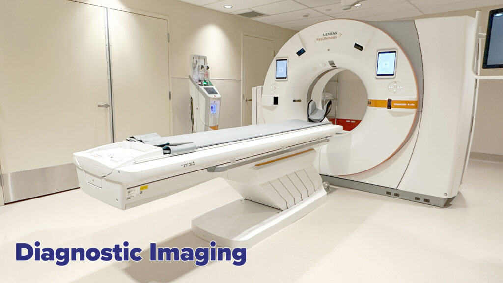
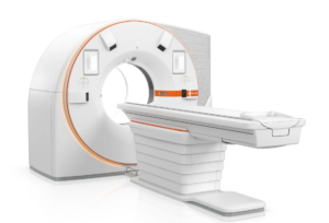 Computed Tomography (CT) scanning is a highly specialized x-ray imaging procedure used to acquire detailed images of every area in the body, providing information no other imaging device can obtain. Modern CT Scanners are an incredibly valuable tool that have revolutionized the diagnosis and treatment of trauma, cancer, stroke, infection and many other conditions.
The Diagnostic Imaging Department at the BCHS is one of the busiest in the region, operating 24/7 and performing 50,000 CT scans every year. 45% of the volume on the CT results from Emergency Department patient visits.
Since the installation of the BCHS’s first CT in 2010, volume/demand increased by 200%. This excessive demand increased wear and tear on the CT unit, resulting in increased downtime. Included in the BCHS’s Emergency Department redevelopment was the purchase of a critically needed second CT Scanner.
